Radiological Imaging Techniques Students Undergo Training on CT Scan Device
On Thursday, October 9, 2025, the Department of Radiological Imaging Techniques at Cihan University-Erbil organized a scientific visit for its fourth-year students to Erbil International Hospital, supervised by Assistant Lecturer Naveed Karimi.
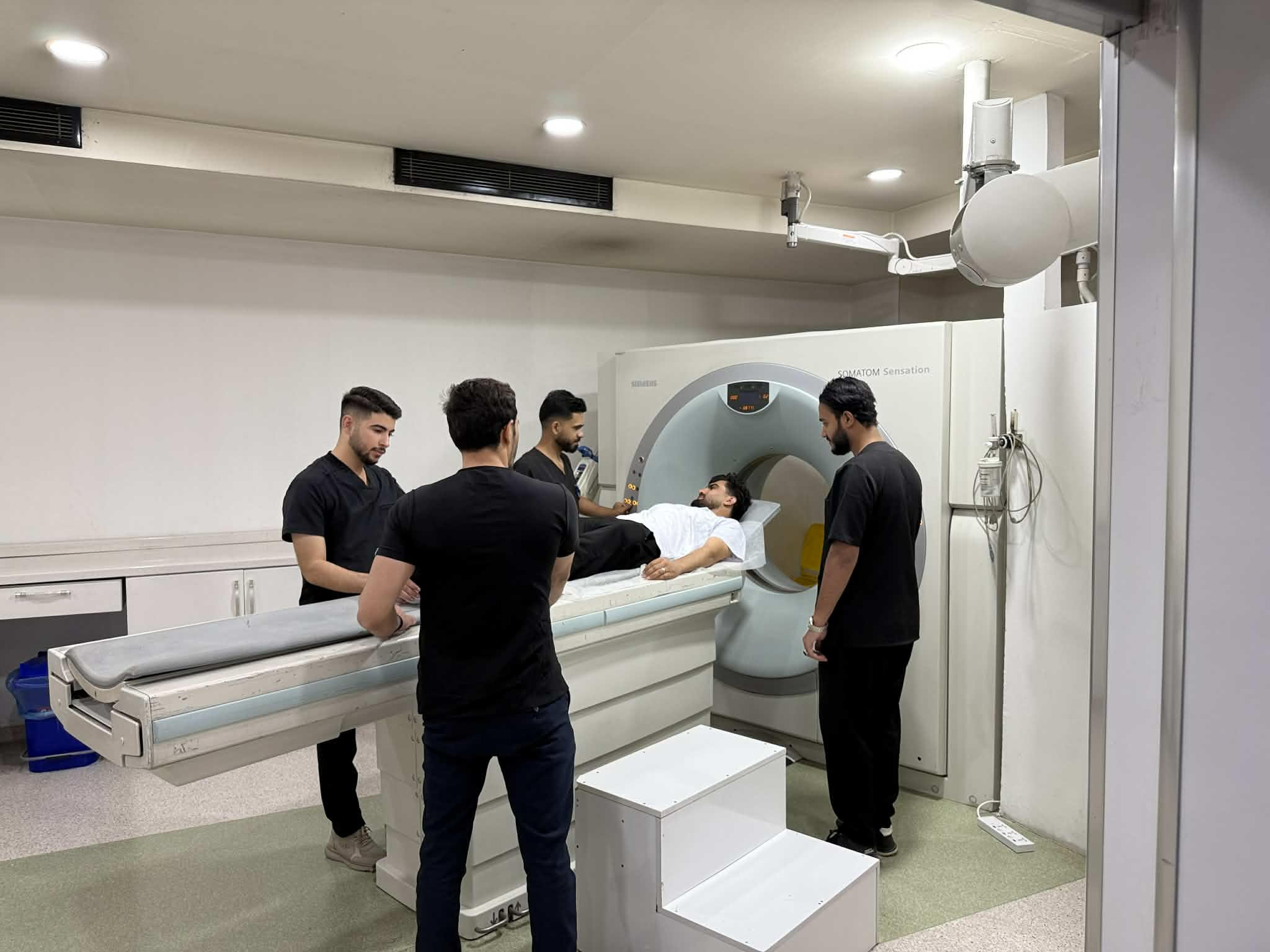
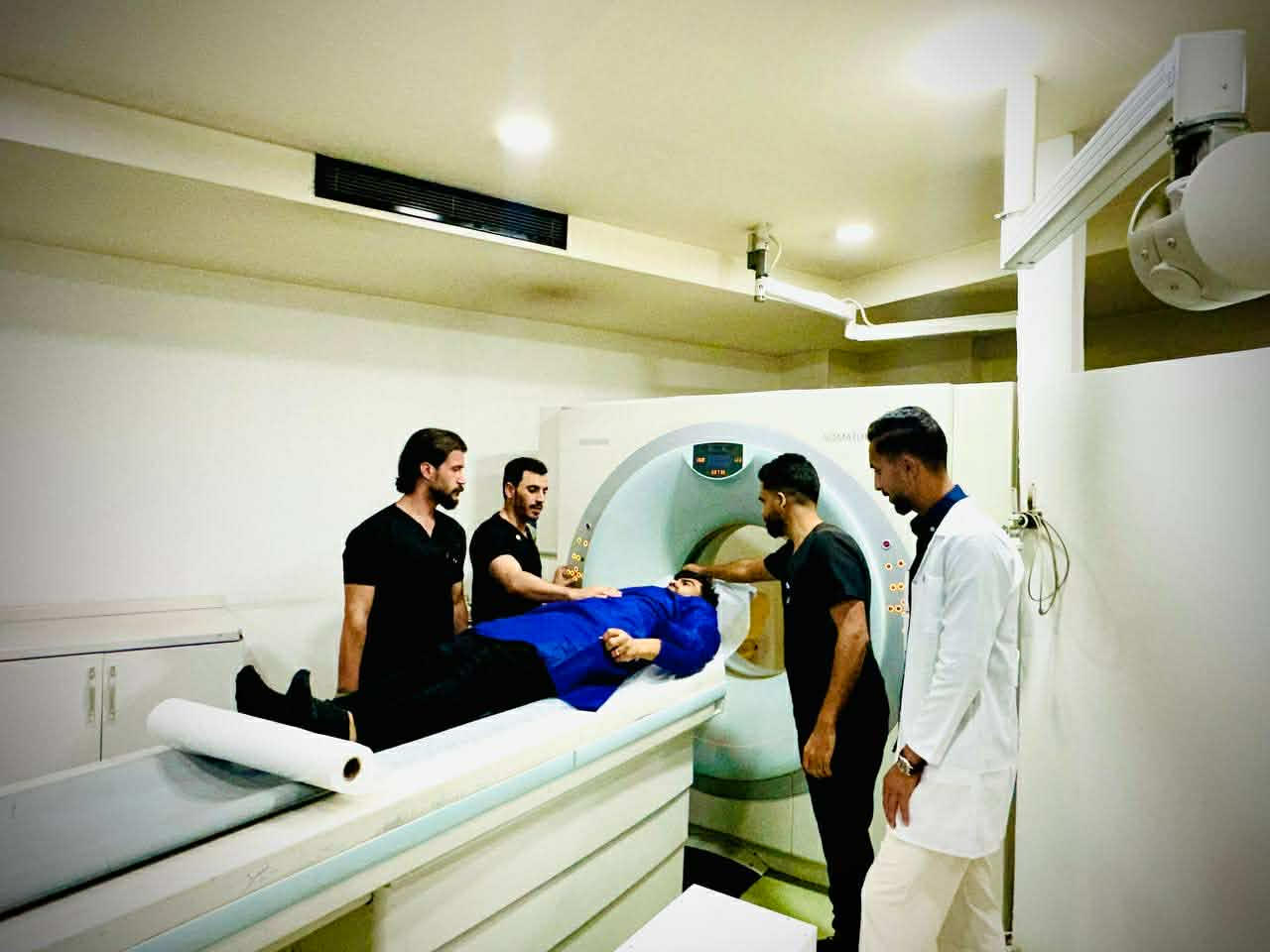
Upon arrival, Ms. Gulala Abdulwahid, a radiologist at the hospital, received the students and provided an overview of the Computed Tomography (CT) Scan device, emphasizing its vital role in diagnosing various diseases. With the assistance of the supervising lecturer, the students then began hands-on training using a Siemens SOMATOM Sensation CT scanner.
Ms. Gulala introduced the students to the main components of the device, explaining its core functions, including the X-ray tube, multi-slice detectors, and control unit. She also guided the students on operating the system, focusing on adjusting key technical parameters such as tube voltage (kVp), current intensity (mA), slice thickness, and spiral rotation speed.
During the session, both the supervising lecturer and the hospital radiologist instructed the students on patient preparation procedures prior to examination. This included assessing the patient’s clinical condition, removing metallic objects that could interfere with image quality, and correctly positioning the patient on the examination table using laser indicators for accurate alignment. The students were also trained to select appropriate imaging modes based on the examination area—such as the brain, chest, abdomen, or pelvis while adhering to the ALARA (As Low As Reasonably Achievable) principle to minimize radiation exposure.
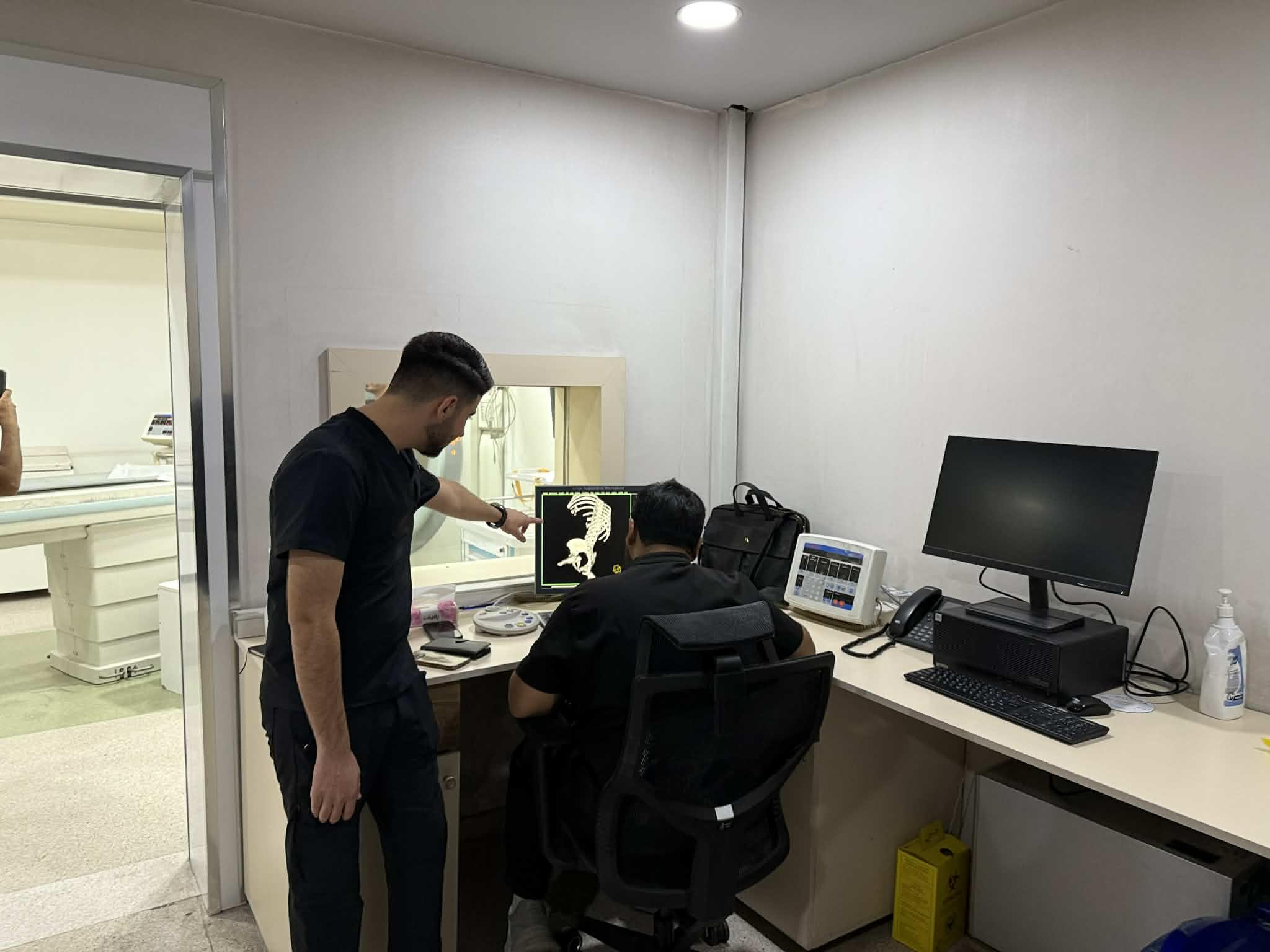
In another part of the visit, the students observed an actual patient imaging procedure in the CT scan room. Afterwards, they proceeded to the control room to review and analyze the obtained images using the digital image processing system.